Home » Without Label » Pelvic Anatomy - Female Pelvic Anatomy Medical Illustration Medivisuals - • divided into the true and false pelvis by the iliopectineal line.
Pelvic Anatomy - Female Pelvic Anatomy Medical Illustration Medivisuals - • divided into the true and false pelvis by the iliopectineal line.
Pelvic Anatomy - Female Pelvic Anatomy Medical Illustration Medivisuals - • divided into the true and false pelvis by the iliopectineal line.. A pelvic ultrasound allows quick visualization of the female pelvic organs and structures including the uterus, cervix, vagina, fallopian tubes and ovaries. Concept for study of anatom. The pelvis is the lower portion of the trunk, located between the abdomen and the lower limbs. Sacrum (the large triangular bone at the base of the spine) The pelvic girdle is the ring shaped collection of these bones at the base of the spine.
Average 3.7 of 19 ratings. List the arterial & nerve supply list the lymph & venous drainage of the pelvis. Pelvic pain can be a sign that there might be a problem with one of the reproductive organs in a woman's pelvic area. Anatomy of female pelvic area. The pelvis is a basin shaped bony structure formed by the combination of two pelvic bones (hip bones or innominate bones) and the sacrum.
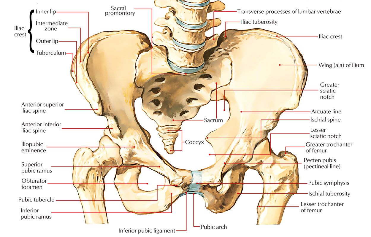
Easy Notes On 【Pelvic Girdle - Coxal Bones】Learn in Just 4 ... from www.earthslab.com The pelvis marks an important transition point between the thoracoabdominal region and the lower limbs.not only is it important for walking, but it also houses organs of the urogenital and distal digestive systems and acts as a conduit for arteries, veins, lymphatic vessels, and nerves necessary for daily functioning. It is usually divided into two separate anatomic regions: A full understanding of pelvic anatomy is required to treat pelvic fractures, to prevent iatrogenic injuries, and to provide the best results. The pelvis is the part of the body located between the abdomen and the thighs. This mri male pelvis axial cross sectional anatomy tool is absolutely free to use. Reproduction system pelvis female woman reproductive system pelvic floor women female bladder and urethra female pelvic floor pelvis woman pelvic floor health woman incontinence pelvis muscles. Reproduction system anatomy isolated on white background. Laparoscopic anatomy of the female pelvic region.
Describe the anatomy of the pelvic wall, bones, joints & muscles.
The pelvis is a musculoskeletal structure that is made up of. The pelvis is the lower part of the torso. A pelvic ultrasound allows quick visualization of the female pelvic organs and structures including the uterus, cervix, vagina, fallopian tubes and ovaries. The pelvic region is the area between the trunk and the lower extremities, or legs. It provides attachment to some important muscles in the region, and forms a cavity which accommodates several important internal organs. The pelvis's frame is made up of the bones of the pelvis, which connect the axial skeleton to the femurs, and therefore acts in weight bearing of the upper body. Sacrum (the large triangular bone at the base of the spine) Female pelvic anatomy what is pelvic pain? Use the mouse scroll wheel to move the images up and down alternatively use the tiny arrows (>>) on both side of the image to move the images.>>) on both side of the image to move the images. It's located between the abdomen and the legs. The pelvic girdle is the ring shaped collection of these bones at the base of the spine. The main function of the pelvic floor musclesare: The pelvic girdle and pelvic spine.
Learning pelvic anatomy is composed of learning bones, muscles, ligaments, nerves and vascular supply. Pelvic hip the hip bone is formed by three bones; A pelvic ultrasound allows quick visualization of the female pelvic organs and structures including the uterus, cervix, vagina, fallopian tubes and ovaries. The pelvic bones are smaller and narrower. This cavity is located within the lesser part of the pelvis, beneath the pelvic brim.
5 Facts about the Anatomy of the Pelvic Cavity from www.visiblebody.com The pelvis's frame is made up of the bones of the pelvis, which connect the axial skeleton to the femurs, and therefore acts in weight bearing of the upper body. The pelvic girdle and pelvic spine. Concept for study of anatom. The term `pelvis` can refer to the pelvic skeleton (also known as the pelvic girdle), which is the skeleton embedded in the lower part of the trunk, connecting the axial skeleton to the lower extremities. Describe the anatomy of the pelvic wall, bones, joints & muscles. Anatomy the pelvis is a ring of bones located at the lower end of the trunk—between the spine and the legs. Differentiate the different types of the female pelvis. The main function of the pelvic floor musclesare:
Reproduction system pelvis female woman reproductive system pelvic floor women female bladder and urethra female pelvic floor pelvis woman pelvic floor health woman incontinence pelvis muscles.
The pelvis opens superiorly to the abdomen through the pelvic inlet, while its inferior opening (the pelvic outlet) is closed by the pelvic floor (levator ani and coccygeus muscles). The pelvic girdle and pelvic spine. Differentiate the different types of the female pelvis. It provides attachment to some important muscles in the region, and forms a cavity which accommodates several important internal organs. The pelvis's frame is made up of the bones of the pelvis, which connect the axial skeleton to the femurs, and therefore acts in weight bearing of the upper body. It is usually divided into two separate anatomic regions: The male pelvis is different from a female's. Average 3.7 of 19 ratings. This cavity is located within the lesser part of the pelvis, beneath the pelvic brim. The pubic bone, also known as pubis, is located inferiorly on the pelvic girdle. Female pelvic anatomy what is pelvic pain? Describe the components & function of the pelvic diaphragm. Describe the boundaries and subdivisions of the pelvis.
The male pelvis is different from a female's. Pubic bones vary in size and shape, but are smaller than the hip bones and form upside down. The pelvis is the lower part of the torso. The lining of the uterus. Ultrasound uses a transducer that sends out.
Pelvic Anatomy Bones from cdn4.vectorstock.com A pelvic ultrasound is a noninvasive diagnostic exam that produces images that are used to assess organs and structures within the female pelvis. This mri male pelvis axial cross sectional anatomy tool is absolutely free to use. It's located between the abdomen and the legs. It provides attachment to some important muscles in the region, and forms a cavity which accommodates several important internal organs. This cavity is located within the lesser part of the pelvis, beneath the pelvic brim. The pelvis is the part of the body located between the abdomen and the thighs. Reproduction system anatomy isolated on white background. Differentiate the different types of the female pelvis.
The pelvic girdle and pelvic spine.
The photo of pelvic is on the woman `s body, isolate on white background, female anatomy concept. Pelvic hip the hip bone is formed by three bones; Differentiate the different types of the female pelvis. Ct body (lymph nodes) ct. Describe the components & function of the pelvic diaphragm. The main function of the pelvic floor musclesare: This cavity is located within the lesser part of the pelvis, beneath the pelvic brim. It's located between the abdomen and the legs. Ultrasound uses a transducer that sends out. Pelvic pain can be a sign that there might be a problem with one of the reproductive organs in a woman's pelvic area. The pelvis's frame is made up of the bones of the pelvis, which connect the axial skeleton to the femurs, and therefore acts in weight bearing of the upper body. The pelvis is the lower portion of the trunk, located between the abdomen and the lower limbs. Reproduction system pelvis female woman reproductive system pelvic floor women female bladder and urethra female pelvic floor pelvis woman pelvic floor health woman incontinence pelvis muscles.